 |
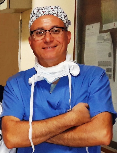
CIRO RUGGIERO
Docente a contratto
Dipartimento di Scienze Mediche e Chirurgiche Materno-Infantili e dell'Adulto
|
Home |
Curriculum(pdf) |
Pubblicazioni
2021
- Use of Octreotide in association with talc poudrage for the management of a severe chylothorax: A case report
[Articolo su rivista]
Lovati, Eleonora; Ruggiero, Ciro; Masciale, Valentina; Stefani, Alessandro; Morandi, Uliano; Aramini, Beatrice
abstract
Chylothorax is an uncommon form of pleural effusion characterized by the presence of chylomicrons, triglycerides and cholesterol in the physical and chemical examination of the pleural fluid. It may have poor prognosis if not properly treated. Currently, conservative measures are the first line of treatment for managing chylothorax. The aim of our study is to show and suggest the use of octreotide in association with talc poudrage as good option to manage post-operative a severe chylothorax.
2020
- Surgery for elastofibroma dorsi: optimizing the management of a benign tumor – an analysis of 70 cases
[Articolo su rivista]
Scamporlino, A; Ruggiero, C; Aramini, B; Morandi, U; Stefani, A
abstract
Background: Elastofibroma dorsi (ED) is a benign soft-tissue tumor of the chest wall located near the
tip of the scapula. Clinical presentation includes swelling, pain and impairment of shoulder movements.
The present literature relies only on few small case series. The aim of this study was to analyze the surgical
management of ED, focusing on the debated topics regarding preoperative evaluation, operative technique,
post-operative outcome and follow-up.
Methods: We conducted a single-center retrospective cohort analysis of patients operated for ED between
2003 and 2018. Diagnostic techniques were ultrasonography (US), computed tomography (CT-scan) and
magnetic resonance imaging (MRI). CT-scan represented our preferred imaging study for preoperative
assessment. Surgery was proposed for symptomatic and/or large lesions. Marginal excision through a musclesparing
approach was performed. An open-door follow-up policy was adopted. All clinical, radiological,
perioperative and pathological variables were matched in a univariate analysis. A multivariate analysis
was performed to investigate risk factors for postoperative complications. Correlations analysis between
radiological and pathological measurements of elastofibroma was conducted.
Results: Seventy elastofibromas were excised in 59 patients. Mean age was 59 years and female prevalence
was 59%. All elastofibromas were completely resected with no recurrence. Postoperative complications
rate was 17%. Complications were mild in most cases. At the univariate analysis, patients with body mass
index (BMI) >25 had a longer operative time (P=0.048), patients on antiplatelet medications experienced
a prolonged drainage time (P=0.006) and a higher rate of complications (P=0.038); the occurrence of
complications resulted in prolonged drainage time (P=0.047) and length of stay (P=0.023). A BMI ≤25 was
the only independent risk factor for postoperative morbidity (OR 8.71, P=0.024). CT-scan showed the
highest correlation with pathological size (r=0.819), US the lowest (r=0.421).
Conclusions: Marginal resection through a muscle-sparing approach is safe and effective for the treatment
of ED. CT-scan can be adequate for preoperative assessment. Giving the benign nature of the lesion and the
absence of recurrence after complete resection, an open-door follow-up may be appropriate.
2020
- Wound complication after modified Ravitch for pectus excavatum: A case of conservative treatment enhanced by pectoralis muscle transposition
[Articolo su rivista]
Aramini, B; Morandi, U; De Santis, G; Brugioni, L; Stefani, A; Ruggiero, C; Baccarani, A
abstract
Ravitcha b s t r a c tINTRODUCTION: Multiple surgical debridement sessions are mandatory before wound closure in cases ofinfection after a modified Ravitch procedure for pectus excavatum. Vacuum-assisted closure (VAC) is awell-established technical resource for treating complicated wounds; however, in cases of suspicion ofbone infection, this approach is not enough to prevent bar removal.PRESENTATION OF THE CASE: We present a case of surgical wound dehiscence with hardware exposure in apatient who had undergone chondrosternoplasty for pectus excavatum. Several sessions of debridement(three) and VAC were applied every time. The final result was achieved without the necessity to removethe hardware; however, to avoid the risk of infection, a bilateral pectoralis muscle flap mobilization wasperformed as the final step after the surgical wound revisions, although this approach is suggested tobe used during the modified Ravitch procedure. This approach allows for a significant reduction in latecomplications and improves morphological outcomes.DISCUSSION: In summary, the pectoralis muscle flap transposition is very useful not only for aestheticalresults but also in combination with multiple surgical revisions for conservative management in caseof wound infection during a modified Ravitch procedure. In our case, this technique was adopted afteraccurate care of the wound and before the final closure, which helps to maintain good vascularizationand a very satisfying result.CONCLUSION: It is important to consider this approach during the modified Ravitch procedure, not onlyfor better aesthetical results but also to prevent infections or wound dehiscence at the level of the bar
2019
- Giant bulla or pneumothorax: How to distinguish.
[Articolo su rivista]
Aramini, Beatrice; Ruggiero, Ciro; Stefani, Alessandro; Morandi, Uliano
abstract
BACKGROUND: The differential diagnosis between pneumothorax and giant bullae is thought to be straightforward but sometimes poses a challenge. CASE PRESENTATION: We present a case of a 54-year-old Caucasian man with a giant emphysematous bulla who underwent surgical resection. He had no smoking history and had previous pneumonia episodes. The surgery was free of complications, without air leaks, and he showed good ventilation of the lung. DISCUSSION: The main complications of bullae are pneumothorax, infection and hemorrhage. Pneumothorax is a serious complication in patients with compromised lung function. Therefore, it is very important to carefully distinguish bullae from pneumothorax to avoid iatrogenic pneumothorax in patients with bullous disease. CONCLUSION: We emphasize how to differentiate between giant bullae and pneumothorax utilizing history, physical examination, and radiological studies, including computed tomography (CT) scan.
2019
- Pectoralis Muscle Transposition in Association with the Ravitch Procedure in the Management of Severe Pectus Excavatum.
[Articolo su rivista]
BACCARANI, ALESSIO; Aramini, Beatrice; DELLA CASA, GIOVANNI; BANCHELLI, FEDERICO; D'AMICO, Roberto; RUGGIERO, Ciro; Starnoni, Marta; Pedone, Antonio; STEFANI, Alessandro; MORANDI, Uliano; DE SANTIS, Giorgio
abstract
Background: Pectus excavatum (PE) is the most common congenital chest wall deformity. PE is sometimes associated with cardiorespiratory impairment, but is often associated with psychological distress, especially for patients in their teenage years. Surgical repair of pectus deformities has been shown to improve both physical limitations and psychosocial well-being in children. The most common surgical approaches for PE treatment are the modified Ravitch technique and the minimally invasive Nuss technique. A technical modification of the Ravitch procedure, which includes bilateral mobilization and midline transposition of the pectoralis muscle flap, is presented here. Methods: From 2010 to 2016, 12 patients were treated by a modified Ravitch procedure with bilateral mobilization and midline transposition of the pectoralis muscle flap for severe PE. Outcomes, morphological results, and complications were analyzed with respect to this new combined surgical approach. Results: There was a statistically significant difference between pre- and postoperative values (P = 0.0025) of the Haller index at the 18-month follow-up, showing a significant morphological improvement for all treated patients. After surgery, no morbidity and mortality were noted. The mean hospital stay was 7 days, and all patients were discharged without major complications. Conclusion: This technique significantly improved patients’ postoperative morphological outcomes and significantly reduced long-term complications, such as wound dehiscence, skin thinning, and hardware exposure.
2018
- An unusual drain in the pleural cavity: iatrogenic pneumothorax due to pulmonary misplacement of a nasogastric tube
[Articolo su rivista]
Stefani, A; Ruggiero, C; Aramini, B; Scamporlino, A
abstract
Gli autori descrivono un raro caso di pneumotorace conseguente a un errato posizionamento intrabronchiale di un sondino nasogastrico in una paziente in stato soporoso
2007
- La chirurgia toracica a Modena. 30 anni di esperienze vissute e di risultati conseguiti
[Articolo su rivista]
Morandi, U; Fontana, G; Lavini, C; Ruggiero, Ciro; Stefani, A; Casali, C; Natali, P; Brandi, L; Chiapponi, A; Lodi, R.
abstract
L'articolo riporta i risultati conseguiti in 30 anni di attività della clinica di chirurgia toracica di modena